ATLAS OF RADIOLOGICAL IMAGES v.1
General University Hospital and 1st Faculty of Medicine of Charles University in Prague
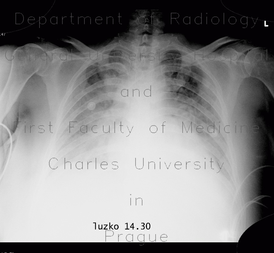
Pumonary edema, pneumonia
CASE
Supine plain chest radiograph shows decreased transparency of both lung wings most pronounced in perihilar regions, consistent with pulmonary edema. CT confirms this finding - multiple areas of consolidation with gravity-dependent density, outlined posteriorly by interlobular septa. CT also shows areas of consolidation in both lower lung lobes consistent with pneumonia and bilateral pleural effusion.


