Renal carcinoma, Grawitz tumour

CASE

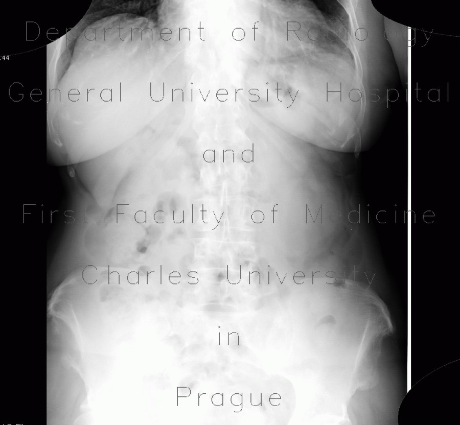
A large mass projection to the lower half of the left kidney can already be appreciated on plain radiograph. CT shows large mass arising from the lower segment of the left kidney that has hypodense (necrotic) center and thick enhancing rim. In correlation, ultrasound shows the rim as hyperechoic and the centre as hypoechoic.

Radiology image - Renal carcinoma, Grawitz tumour: Abdomen, Kidney and adrenals: X-ray - Plain radiograph

Renal carcinoma, Grawitz tumour


Renal carcinoma, Grawitz tumour: X-ray - Plain radiographRenal carcinoma, Grawitz tumour: X-ray - Plain radiographRenal carcinoma, Grawitz tumour: CT - Computed tomographyRenal carcinoma, Grawitz tumour: CT - Computed tomographyRenal carcinoma, Grawitz tumour: CT - Computed tomographyRenal carcinoma, Grawitz tumour: CT - Computed tomographyRenal carcinoma, Grawitz tumour: CT - Computed tomographyRenal carcinoma, Grawitz tumour: CT - Computed tomographyRenal carcinoma, Grawitz tumour: CT - Computed tomographyRenal carcinoma, Grawitz tumour: US - UltrasoundRenal carcinoma, Grawitz tumour: US - Ultrasound
lights on off