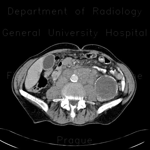
Renal carcinoma, cystic, retroperitoneal lymphadenopathy

CASE

A large cystic formation, which arises from the lower segment of the left kidney, has partially calcified wall, but, what matters most, several enhancing soft tissue nodules in the wall. Note also retroperitoneal lymphadenopathy.

Radiology image - Renal carcinoma, cystic, retroperitoneal lymphadenopathy: Abdomen, Kidney and adrenals: CT - Computed tomography

Renal carcinoma, cystic, retroperitoneal lymphadenopathy


Renal carcinoma, cystic, retroperitoneal lymphadenopathy: CT - Computed tomographyRenal carcinoma, cystic, retroperitoneal lymphadenopathy: CT - Computed tomographyRenal carcinoma, cystic, retroperitoneal lymphadenopathy: CT - Computed tomographyRenal carcinoma, cystic, retroperitoneal lymphadenopathy: CT - Computed tomographyRenal carcinoma, cystic, retroperitoneal lymphadenopathy: CT - Computed tomographyRenal carcinoma, cystic, retroperitoneal lymphadenopathy: CT - Computed tomographyRenal carcinoma, cystic, retroperitoneal lymphadenopathy: CT - Computed tomography
lights on off