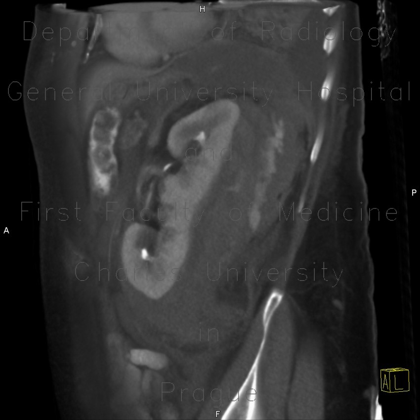
Renal hemorrhage, staghorn calculus

CASE

A thick hypodense sac around the left kidney in the perirenal space with signs of active extravasation of opacified blood (with contrast). On ultrasound, this appears as formation of mixed echogenicity around the left kidney with hypoechoic centre which is caused by fresh blood. Right kidney contains a staghorn calculus.

Radiology image - Renal hemorrhage, staghorn calculus: Abdomen, Kidney and adrenals: CT - Computed tomography

Renal hemorrhage, staghorn calculus


Renal hemorrhage, staghorn calculus: CT - Computed tomographyRenal hemorrhage, staghorn calculus: CT - Computed tomographyRenal hemorrhage, staghorn calculus: CT - Computed tomographyRenal hemorrhage, staghorn calculus: CT - Computed tomographyRenal hemorrhage, staghorn calculus: CT - Computed tomographyRenal hemorrhage, staghorn calculus: CT - Computed tomographyRenal hemorrhage, staghorn calculus: CT - Computed tomographyRenal hemorrhage, staghorn calculus: CT - Computed tomographyRenal hemorrhage, staghorn calculus: CT - Computed tomographyRenal hemorrhage, staghorn calculus: CT - Computed tomographyRenal hemorrhage, staghorn calculus: CT - Computed tomographyRenal hemorrhage, staghorn calculus: CT - Computed tomographyRenal hemorrhage, staghorn calculus, VRT: CT - Computed tomographyRenal hemorrhage, staghorn calculus, VRT: CT - Computed tomographyRenal hemorrhage, staghorn calculus: US - UltrasoundRenal hemorrhage, staghorn calculus: US - UltrasoundRenal hemorrhage, staghorn calculus: US - UltrasoundRenal hemorrhage, staghorn calculus: US - Ultrasound
lights on off