Rupture of the diaphragm, diaphragmatic hernia

CASE

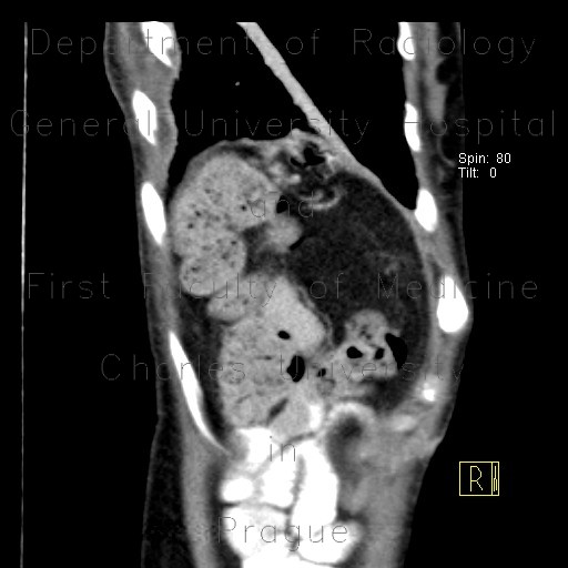
CT shows a prolapse of abdominal viscera into the left pleural cavity through the ruptured left hemidiaphragm. The intestinal bowel loops with mixed content can be readily recognized in the chest radiograph.

Radiology image - Rupture of the diaphragm, diaphragmatic hernia: Abdomen, Thorax, Large bowel, Mediastinum and pleural cavity, Peritoneal cavity, Soft tissue: CT - Computed tomography

Rupture of the diaphragm, diaphragmatic hernia


Rupture of the diaphragm, diaphragmatic hernia: CT - Computed tomographyRupture of the diaphragm, diaphragmatic hernia: X-ray - Plain radiographRupture of the diaphragm, diaphragmatic hernia: X-ray - Plain radiographRupture of the diaphragm, diaphragmatic hernia: CT - Computed tomographyRupture of the diaphragm, diaphragmatic hernia: CT - Computed tomographyRupture of the diaphragm, diaphragmatic hernia: CT - Computed tomographyRupture of the diaphragm, diaphragmatic hernia: CT - Computed tomographyRupture of the diaphragm, diaphragmatic hernia: CT - Computed tomographyRupture of the diaphragm, diaphragmatic hernia: CT - Computed tomographyRupture of the diaphragm, diaphragmatic hernia: CT - Computed tomographyRupture of the diaphragm, diaphragmatic hernia: CT - Computed tomographyRupture of the diaphragm, diaphragmatic hernia: CT - Computed tomographyRupture of the diaphragm, diaphragmatic hernia: CT - Computed tomography
lights on off