Sclerodermia, pulmonary fibrosis, UIP, fibrosis, oesophagus

CASE

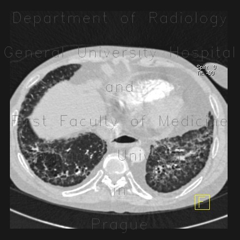
CT of the thorax shows fibrous changes most pronounced in lower lung lobes. These include interlobular septal thickening and traction bronchiectasis. The presence of ground-glass opacity indicates alveolitis in an active disesase. Note also that the lumen of esophagus is widened due to involvement by sclerodermia.

Tags:  Thorax  Lung  Oesophagus
Radiology image - Sclerodermia, pulmonary fibrosis, UIP, fibrosis, oesophagus: Thorax, Lung, Oesophagus: CT - Computed tomography

Sclerodermia, pulmonary fibrosis, UIP, fibrosis, oesophagus


Sclerodermia, pulmonary fibrosis, UIP, fibrosis, oesophagus: CT - Computed tomographySclerodermia, pulmonary fibrosis, UIP, fibrosis, oesophagus: CT - Computed tomographySclerodermia, pulmonary fibrosis, UIP, fibrosis, oesophagus: CT - Computed tomographySclerodermia, pulmonary fibrosis, UIP, fibrosis, oesophagus: CT - Computed tomographySclerodermia, pulmonary fibrosis, UIP, fibrosis, oesophagus: CT - Computed tomographySclerodermia, pulmonary fibrosis, UIP, fibrosis, oesophagus: CT - Computed tomographySclerodermia, pulmonary fibrosis, UIP, fibrosis, oesophagus: CT - Computed tomography
lights on off