ATLAS OF RADIOLOGICAL IMAGES v.1
General University Hospital and 1st Faculty of Medicine of Charles University in Prague
Sequestrum, osteomyelitis of upper jaw, maxilla
CASE
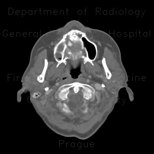
This patient developed a dentigennous osteomyelitis of the right part of the maxilla. CT shows involvement of the bony palate with a bony defect that contains a small sequestrum. The bone itself is thickened (compared to the left half of the maxilla) and the right maxillary sinus is filled with markedly thickened mucosa filling almost entire cavity.


