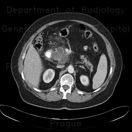
Serous adenoma of pancreas, microcystic

CASE

CT shows a septated cystic mass in the head of pancreas. It has thin septa that show mild enhancement and the cystic spaces are smaller than 1cm. Pancreatic duct is slightly dilated. Ultrasound shows a cystic formation in the head of pancreas but its structure can not be discerned due to worse quality of the image in an obese patient.

Tags:  Abdomen  Pancreas
Radiology image - Serous adenoma of pancreas, microcystic: Abdomen, Pancreas: CT - Computed tomography

Serous adenoma of pancreas, microcystic


Serous adenoma of pancreas, microcystic: CT - Computed tomographySerous adenoma of pancreas, microcystic: CT - Computed tomographySerous adenoma of pancreas, microcystic: CT - Computed tomographySerous adenoma of pancreas, microcystic: CT - Computed tomographySerous adenoma of pancreas, microcystic: CT - Computed tomographySerous adenoma of pancreas, microcystic: CT - Computed tomographySerous adenoma of pancreas, microcystic: CT - Computed tomographySerous adenoma of pancreas, microcystic: CT - Computed tomographySerous adenoma of pancreas, microcystic: CT - Computed tomographySerous adenoma of pancreas, microcystic: CT - Computed tomographySerous adenoma of pancreas, microcystic: CT - Computed tomographySerous adenoma of pancreas, microcystic: CT - Computed tomographySerous adenoma of pancreas, microcystic: US - Ultrasound
lights on off