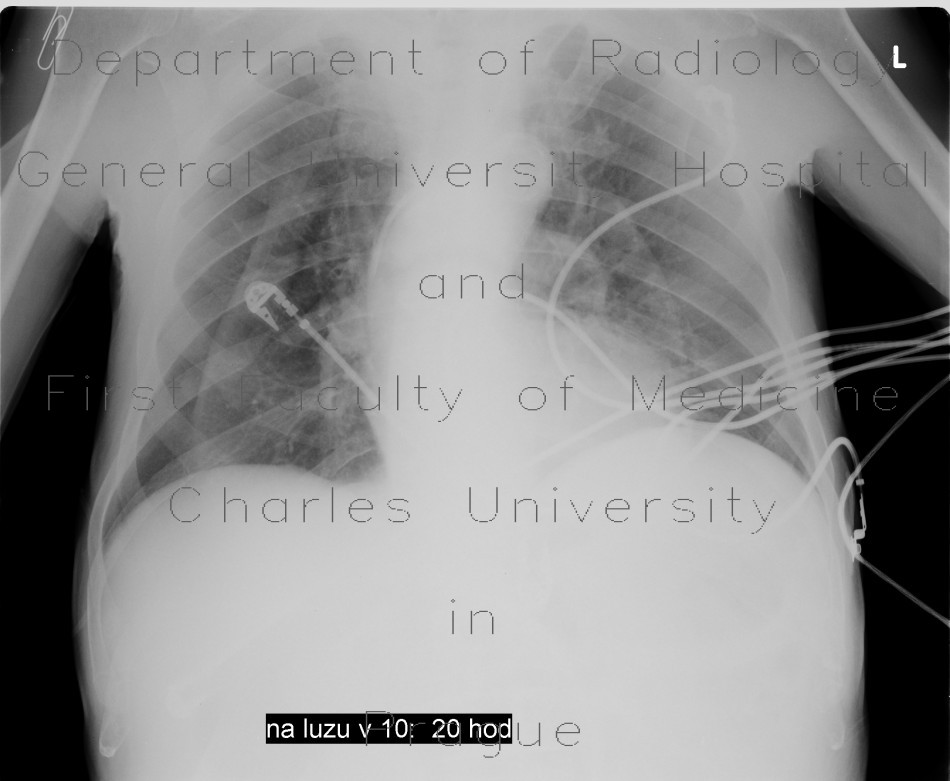
Skin folds, mimic of pneumothorax, PNO

CASE

Chest radiograph shows a curved line which creates a border between normal transparency in the periphery and a gradient of increased opacity that fades toward the mediastinum. Vascular markings are visible beyond these lines on both sides, pneumothorax can be safely excluded. CT shows how skin folds are formed in supine position.

Radiology image - Skin folds, mimic of pneumothorax, PNO: Thorax, Lung, Mediastinum and pleural cavity: X-ray - Plain radiograph

Skin folds, mimic of pneumothorax, PNO


Skin folds, mimic of pneumothorax, PNO: X-ray - Plain radiographSkin folds, mimic of pneumothorax, PNO: CT - Computed tomographySkin folds, mimic of pneumothorax, PNO: CT - Computed tomographySkin folds, mimic of pneumothorax, PNO: CT - Computed tomographySkin folds, mimic of pneumothorax, PNO: CT - Computed tomographySkin folds, mimic of pneumothorax, PNO: CT - Computed tomographySkin folds, mimic of pneumothorax, PNO: CT - Computed tomographySkin folds, mimic of pneumothorax, PNO: CT - Computed tomography
lights on off