ATLAS OF RADIOLOGICAL IMAGES v.1
General University Hospital and 1st Faculty of Medicine of Charles University in Prague
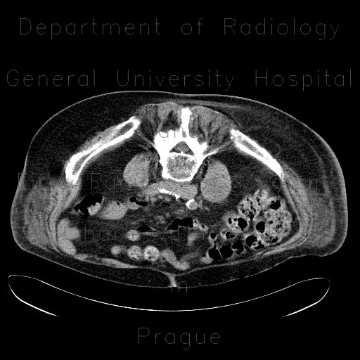
Spondylodiscitis, abscess of psoas muscle, drainage of abscess
CASE
CT shows a fusiform cavity in the right psoas muscle. The cavity has well-defined wall and contains fluid (CT attenuation, high on T2, low on T1). The L2 and L3 intervertebral discs are replaced by fluid. The abscess was drained under CT guidance.


