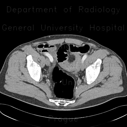
Stenosis of sigmoid colon and lienal flexure, inflammatory stenosis, CT colonography

CASE

CT shows segmental thickening of wall of sigmoid colon with mild edema of adjacent fat and peritoneum. Another stenotic segment can be seen in the lienal flexure, probably due to mild diverticulitis.

Radiology image - Stenosis of sigmoid colon and lienal flexure, inflammatory stenosis, CT colonography: Abdomen, Large bowel: CT - Computed tomography

Stenosis of sigmoid colon and lienal flexure, inflammatory stenosis, CT colonography


Stenosis of sigmoid colon and lienal flexure, inflammatory stenosis, CT colonography: CT - Computed tomographyStenosis of sigmoid colon and lienal flexure, inflammatory stenosis, CT colonography: CT - Computed tomographyStenosis of sigmoid colon and lienal flexure, inflammatory stenosis, CT colonography: CT - Computed tomographyStenosis of sigmoid colon and lienal flexure, inflammatory stenosis, CT colonography: CT - Computed tomographyStenosis of sigmoid colon and lienal flexure, inflammatory stenosis, CT colonography: CT - Computed tomographyStenosis of sigmoid colon and lienal flexure, inflammatory stenosis, CT colonography: CT - Computed tomography
lights on off