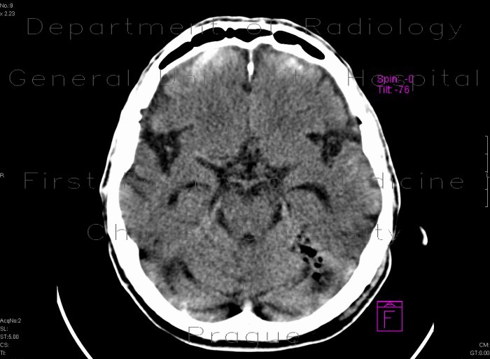
Subarachnoid hemorrhage, beam hardening artifact, pneumocephalus

CASE

Posttraumatic subarachnoid hemorrhage of both frontal pole - do not confuse with mere artifacts, which are also present. Decoloration on follow-up scans. Small bubbles at the insertion of tentorium on the left.

Tags:  Brain  Brain
Radiology image - Subarachnoid hemorrhage, beam hardening artifact, pneumocephalus: Brain, Brain: CT - Computed tomography

Subarachnoid hemorrhage, beam hardening artifact, pneumocephalus


Subarachnoid hemorrhage, beam hardening artifact, pneumocephalus: CT - Computed tomographySubarachnoid hemorrhage, beam hardening artifact, pneumocephalus: CT - Computed tomographySubarachnoid hemorrhage, beam hardening artifact, pneumocephalus: CT - Computed tomographySubarachnoid hemorrhage, beam hardening artifact, pneumocephalus: CT - Computed tomographySubarachnoid hemorrhage, beam hardening artifact, pneumocephalus: CT - Computed tomography
lights on off