Thymoma, compression of the brachiocephalic vein, collateral flow

CASE

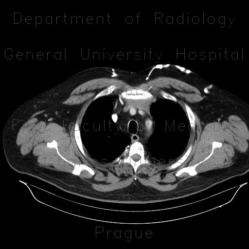
CT shows a soft-tissue mass in the anterior mediastinum that causes compression of the left brachiocephalic vein. The contrast that was injected to the left arm opacifies collateral flow in the left chest.

Radiology image - Thymoma, compression of the brachiocephalic vein, collateral flow: Thorax, Mediastinum and pleural cavity, Vessels: CT - Computed tomography

Thymoma, compression of the brachiocephalic vein, collateral flow


Thymoma, compression of the brachiocephalic vein, collateral flow: CT - Computed tomographyThymoma, compression of the brachiocephalic vein, collateral flow: CT - Computed tomographyThymoma, compression of the brachiocephalic vein, collateral flow: CT - Computed tomographyThymoma, compression of the brachiocephalic vein, collateral flow: CT - Computed tomographyThymoma, compression of the brachiocephalic vein, collateral flow: CT - Computed tomographyThymoma, compression of the brachiocephalic vein, collateral flow: CT - Computed tomographyThymoma, compression of the brachiocephalic vein, collateral flow: CT - Computed tomographyThymoma, compression of the brachiocephalic vein, collateral flow: CT - Computed tomographyThymoma, compression of the brachiocephalic vein, collateral flow: CT - Computed tomography
lights on off