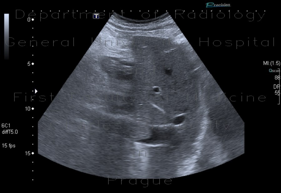
Tumorous thrombosis of inferior vena cava, adrenal metastasis, conventional renal carcinoma

CASE

The ultrasound images show echoic filling of the lumen of the inferior vena cava which representes a thrombus. There is anechoic lumen above it, because the thrombus end in the hepatic portion. Moreover, there is a round expansion in the right adrenal gland representing adrenal metastasis - this cannot be told from ultrasound itself, however.

Radiology image - Tumorous thrombosis of inferior vena cava, adrenal metastasis, conventional renal carcinoma: Abdomen, Kidney and adrenals, Vessels: US - Ultrasound

Tumorous thrombosis of inferior vena cava, adrenal metastasis, conventional renal carcinoma


Tumorous thrombosis of inferior vena cava, adrenal metastasis, conventional renal carcinoma: US - UltrasoundTumorous thrombosis of inferior vena cava, adrenal metastasis, conventional renal carcinoma: US - UltrasoundTumorous thrombosis of inferior vena cava, adrenal metastasis, conventional renal carcinoma: US - UltrasoundTumorous thrombosis of inferior vena cava, adrenal metastasis, conventional renal carcinoma: US - UltrasoundTumorous thrombosis of inferior vena cava, adrenal metastasis, conventional renal carcinoma: US - Ultrasound
lights on off