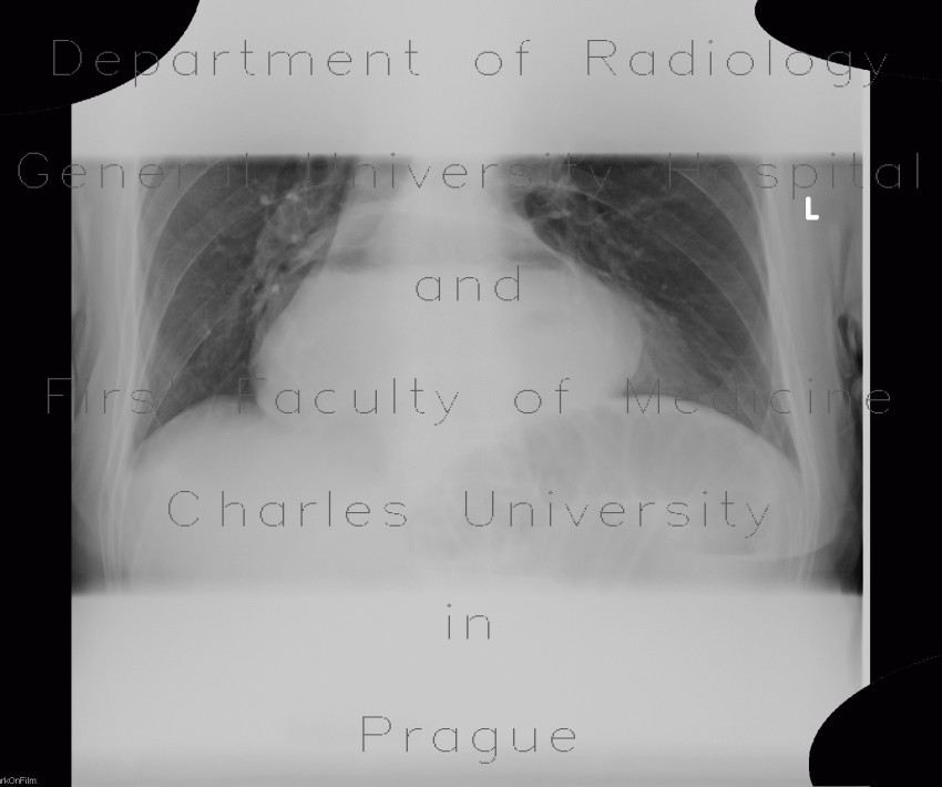
Upside-down stomach, small bowel obstruction, ileus, retained contrast in colonic diverticula

CASE

A large air-fluid level can be readily recognized within the shadow of the heart, CT confirmed an upside-down stomach. Moreover, this patient has small bowel obstruction, which can be recognized on plain radiograph by several dilated small bowel loops with air-fluid levels. CT show the same. Note dense concent of diverticula of the descendent colon - this contrast was retained after previous examination.

Radiology image - Upside-down stomach, small bowel obstruction, ileus: Abdomen, Large bowel, Small bowel, Stomach: X-ray - Plain radiograph

Upside-down stomach, small bowel obstruction, ileus


Upside-down stomach, small bowel obstruction, ileus: X-ray - Plain radiographUpside-down stomach, small bowel obstruction, ileus: X-ray - Plain radiographUpside-down stomach, small bowel obstruction, ileus, retained contrast in colonic diverticula: CT - Computed tomographyUpside-down stomach, small bowel obstruction, ileus, retained contrast in colonic diverticula: CT - Computed tomographyUpside-down stomach, small bowel obstruction, ileus, retained contrast in colonic diverticula: CT - Computed tomographyUpside-down stomach, small bowel obstruction, ileus, retained contrast in colonic diverticula: CT - Computed tomographyUpside-down stomach, small bowel obstruction, ileus, retained contrast in colonic diverticula: CT - Computed tomographyUpside-down stomach, small bowel obstruction, ileus, retained contrast in colonic diverticula: CT - Computed tomographyUpside-down stomach, small bowel obstruction, ileus, retained contrast in colonic diverticula: CT - Computed tomographyUpside-down stomach, small bowel obstruction, ileus, retained contrast in colonic diverticula: CT - Computed tomography
lights on off