ATLAS OF RADIOLOGICAL IMAGES v.1
General University Hospital and 1st Faculty of Medicine of Charles University in Prague
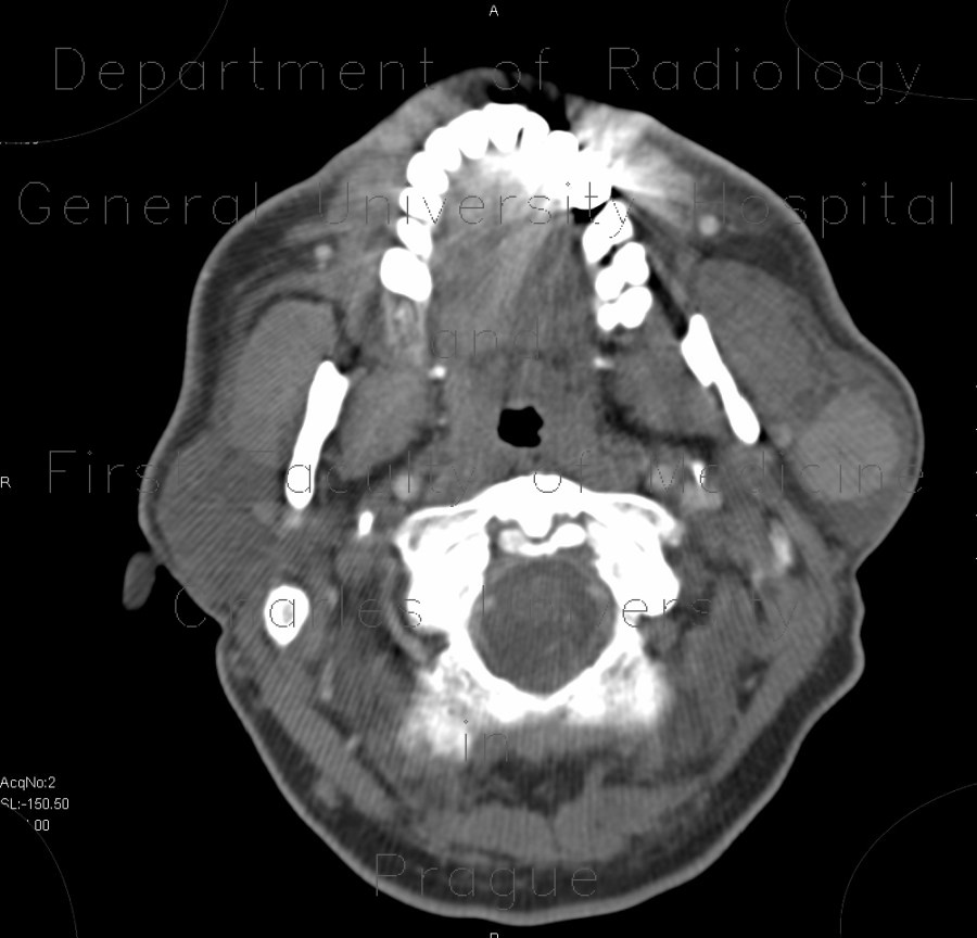
Warthin tumor, parotid gland
CASE
CT shows enhancing well-defined focal lesion in the left parotid gland. In correlation, it has hypoechoic echostructure with some internal echos and perfusion on ultrasound. Histology confirmed Warthin tumor. Warthin tumour and pleomorphic adenoma are most common tumours of the parotid glad. Both are benign, but can grow and in rare cases transform into malignant forms. Their differentiation by ultrasound or CT is very difficult and with very limited confidence.


